|
Diseases of Poultry
By Ivan Dinev, DVM, PhD
|
NECROTIC ENTERITIS
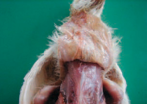
113.. Necrotic enteritis (NE) is an acute Clostridium infection characterized by severe necroses of intestinal mucosa. The disease begins suddenly, with a sharp increase in death rate. A strong dehydration is observed. The skin is sticked on or adhered to body musculature and is hardly removed.
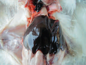
114.Chickens at the age of 25 weeks are usually affected, NE is also encountered in hens particularly near the period of the beginning of egg laying or peak egg laying, most commonly associated with coccidiosis. In acute cases, marked congestion of liver, responsible for its dark red to black appearance, is present.
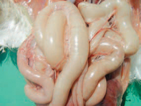
115.The aetiological agent is Clostridium perfringens, mainly from type A and more rarely from type C. The produced a and p" toxins, from C. perfringens type A and type C respectively, are responsible for the necrosis of intestinal mucosa. The small intestine is often distended with gases and the necrotic mucosa is visible through the wall.
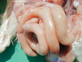
116.CI. perfringens is ubiquitous and normally reside into the intestinal tract. The alterations are particularly in the jejunum and the ileum because of their higher pH and the lower oxygen content in these areas. Sometimes, haemorrhages are seen through the intestinal wall.
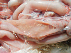
117.The intestinal lumen is filled with brownish watery content, mixed with gas bubbles.
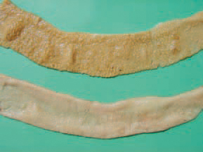
118.necrotic mucosa acquires a greyish-creamy or greenish appearance. Sometimes the mucosa has a flannelette blanket-like appearance.
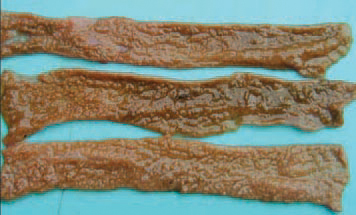
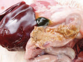
119.120.In some cases, the mucosa has a linear pattern similar to the bark of a tree. The predisposing factors are injuries of intestinal mucosa by various Eimeria species, migration of ascarids, immuno-deficiency states due to CIA, IBD, MD, high content of wheat or fish meal in the diet.
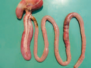
121In cases when NE is associated with small intestinal coccidioses, multiple petechial haemorrhages could be perceived through the wall in different areas along the small intestine.
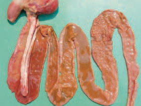
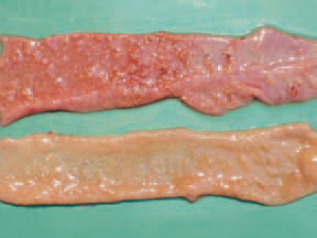
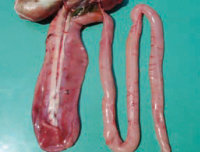
112.123.124.Throughout the simultaneous occurrence of NE and cocci-dioses, the content of the lumen is bloody, mixed with necrotic detritus and gas bubbles. The diagnosis is based on the distinctive gross lesions. When necessary, a histological investigation is performed or attempts for isolation of the causative agent. NE should be distinguished from ulcerative enteritis and some small intestinal cocci-dioses. The control should be aimed at predisposing factors. An appropriate medication of feeds is recommended. A good effect is obtained with oxytetra-cycline dihydrate (OTC 50% premix). NE could be effectively treated with doxy-cycline hydrochloride, amoxicillin etc.


