

Avian Influenza: The role of coinfections
Introduction
Avian Influenza is one of the most important diseases worldwide. It is caused by avian influenza viruses (AIV's) and belongs to the family Orthomyxoviridae, genus Influenza Virus, which has three types, A, B and C. The only Influenza Virus in poultry is type A. The AIV's are classified according to two different pathotypes, Highly Pathogenic Avian Influenza (HPAI) and Low Pathogenic Avian Influenza (LPAI) and any Influenza A virus with an Intravenous Pathogenicity Index greater than 1.2 (or mortality of 75% in four to eight-week-old intravenously infected chicks) is HPAI. According to the World Organisation for Animal Health (OIE), all HPAI, and H5 and H7 cases (regardless of the pathotype) have to be notified. The reason why the LPAI H5 and H7 have to be notified is their potential to become HPAI. The different pathogenicity between LPAI and HPAI is related to the different replication sites in the host. The HPAI can replicate and damage a wide range of organs whilst the LPAI is capable of replicating only in limited tissues of the respiratory and digestive systems (Mo, I. P., 1997). In most cases, LPAI causes asymptomatic or mild infection (Sajid, 2017) but some LPAI viruses cause over 10% mortality.
AI is present on almost every continent, especially Asia, Africa and Europe. It is commonly isolated in wild birds, especially aquatic birds. Control of LPAI in some countries is very difficult, especially because of the reservoirs of aquatic birds.
Coinfection is an infection of an organism by more than one virus or bacterium (Sajid, 2017).
What is the role of coinfections in LPAI?
Several trials have suggested the difficulty of replicating the Avian Influenza virus experimentally (Swayne, 1996), with mild infections often being caused in the trial, suggesting that some other factor is causing the increase in clinical signs.
LPAI coinfections are quite common and normally cause complications of the clinical signs. The infection of a previously infected host can affect the replication with the second infection and the clinical signs. In some cases, there is viral interference, when the primary virus will not permit the replication of a second one. On the other hand, in other cases the replication of the second virus can increase.
The diagnosis in coinfection cases can be difficult in cases of viral interference; in some cases, the virus titres will be low or undetectable.
Two of the most important viral coinfections are LPAI and Infectious Bronchitis and LPAI and Newcastle Disease.
LPAI and Infectious Bronchitis coinfection
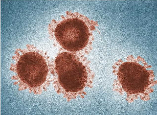
Infectious Bronchitis is an important disease, causing respiratory and productive problems. It is caused by a Coronavirus, and an important characteristic of this virus is one of the structural proteins, the Spike protein (S), the variation in serotypes of the IBV being related to the variation of the S protein (Cavanagh, 1998). Cross-protection from the different serotypes is partial or non-existent. The Massachusetts protectotype (Mass - Classical) is very important and is present worldwide (Sylvester, 2005). The Mass Protectotype has a respiratory tropism.
Normally in the respiratory syndrome, the possibility of the involvement of one or more agents is high (Habibbolah, 2019).
With regard to H9N2 coinfection with the IBV, an experimental study carried out in Iran suggested an increase in clinical signs and a significant increase in the titre against LPAI, indicating the possibility of the IBV promoting the replication of the LPAIV. The classical IB strain (H120) and variants were used in the trial (Seifi, 2010). In one experimental trial in Egypt, coinfection with LPAI and the IBV caused an increase in the shedding of the AIV (Kareen, 2017). This means that the IBV can aggravate clinical signs of AI and accelerate the spread of AI and means that IB should be controlled in LPAI endemic areas.
To prevent IB, it is important to use appropriate IB vaccines that counteract the endemic field strain. Molecular diagnosis (Sequencing) is the key to understanding the prevalent IBV variants in the region.

The use of a classical IB (H120) strain is one important point in helping to control IB and consequently to control the most adverse consequences of LPAI.
LPAI and Newcastle Disease coinfection
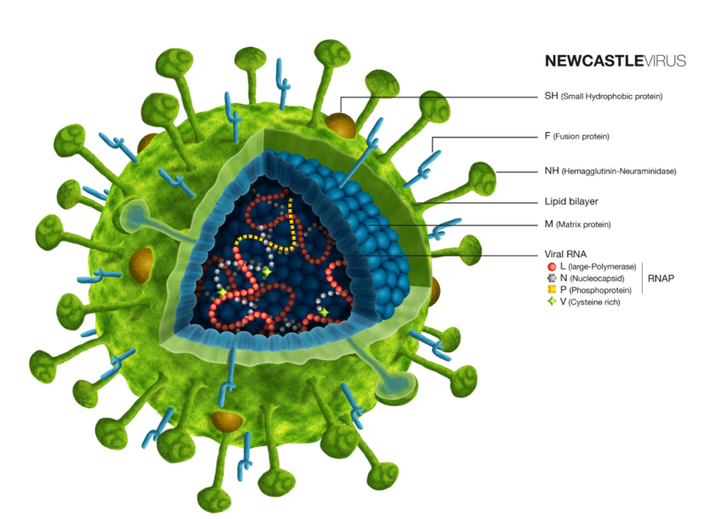
Newcastle disease is present in several countries, causing mortality and economic and trading losses. Newcastle disease is caused by a paramyxovirus, the Avian Paramyxovirus serotype 1 and one important characteristic of this disease is the presence of just one serotype, regardless of the different genotypes.
It is suggested that LPAI and Newcastle disease coinfections lower replication of the Newcastle Disease Virus (NDV). Both NDV and AIV bind to sialic acid–linked glycoconjugates on host cells and may also compete for host cell machinery during viral replication.(Sajid, 2017) Coinfection of LPAI and NDV in ducks resulted in a higher number of positive cloacal swabs detected for LPAIV and a lower number of cloacal swabs detected positive for NDV (Franca, 2014). However, these affected replication dynamics do not change the clinical signs (Mar, 2014)
In this case, the lower replication rate and no changes in clinical signs make diagnosis a problem; in some cases, it is possible that the coinfection interferes with the ND titres.

© Source: HIPRA
In cases of Coinfections with NDV, it is important to be aware of the interference of LPAI and NDV, and as both of these viruses have respiratory tropism, it is important to use ND vaccines with a respiratory tropism to induce local protection, but with a lower pathogenicity.
Conclusions
To control coinfections with LPAI and IB, the key is to know the IB strains present in the region, to use the vaccines according to the IB variants present locally and to make sure of a strong programme of IB Classical strains.
In the case of coinfections with LPAI and NDV, it is important to take into account the possibility of interference by the LPAI.
HIPRA, as The Reference in Prevention for Animal Health has solutions for the control of Newcastle Disease and Classical Infectious Bronchitis.
The links for the Newcastle disease range are below, highlighting the Hipraviar® Clon and Hipraviar® Clon/H120, LaSota cloned vaccines with high titres and low pathogenicy:
https://www.hipra.com/portal/en/hipra/animalhealth/products/detail/hipraviar-clon
https://www.hipra.com/portal/en/hipra/animalhealth/products/detail/hipraviar-clon-h120
https://www.hipra.com/portal/en/hipra/animalhealth/products/detail/hipraviar-b1
https://www.hipra.com/portal/en/hipra/animalhealth/products/detail/hipraviar-b1-h120
https://www.hipra.com/portal/en/hipra/animalhealth/products/detail/hipraviar-s
https://www.hipra.com/portal/en/hipra/animalhealth/products/detail/hipraviar-s-h120
The Link below for the Bronipra® 1, a classical H-120 Infectious Bronchitis vaccine, very important strain in the IB vaccination program:
https://www.hipra.com/portal/en/hipra/animalhealth/products/detail/bronipra-1
| References | ||||
|---|---|---|---|---|
| Mo I. P., M. Brugh, O. J. Fletcher, G. N. Rowland, and D. E. Swayne. | ||||
| (1997) | Comparative pathology of chickens experimentally inoculated with avian influenza viruses of low and high pathogenicity.. Avian Dis. | 41:125–136. 1997. | ||
| Sajid Umar, Jean Luc Guerin, and Mariette F. Ducatez. | ||||
| (2017) | Low Pathogenic Avian Influenza and Coinfecting Pathogens: A Review of Experimental Infections in Avian Models.. Avian Dis. | 61:3–15, 2017. | ||
| Habibbolah Haji-Abdolvahab, Arah Ghalyanchilangeroudi, Alireza Bahonar, Seyed Ali Ghafouri, Mehdi Vasfi Marandi, Mohammad Hosein Fallah Mehrabadi, Farshad Tehrani. | ||||
| (2019) | Prevalence of avian influenza, Newcastle disease, and infectious bronchitis viruses in broiler flocks infected with multifactorial respiratory diseases in Iran, 2015–2016.. Tropical Animal Health and Production. | 51:689–695, 2019. | ||
| França M, Howerth EW, Carter D, Byas A, Poulson R, Afonso CL, et al. | ||||
| (2014) | Co-infection of mallards with low-virulence Newcastle disease virus and low-pathogenic avian influenza virus.. Avian Pathology; | 43(1):96-104, 2014. | ||
| Seifi S., Asasi, K., and Mohammadi, A. | ||||
| (2010) | Natural co-infection caused by avian influenza H9 subtype and infectious bronchitis viruses in broiler chicken farms.. Vet Arhiv. | 80, 269–281., 2010 | ||
| Kareem E. Hassan, Salama A. S. Shany, A. Ali, Al-Hussien M. Dahshan, Azza A. El-Sawah, and Magdy F. El-Kady. | ||||
| (2016) | Prevalence of avian respiratory viruses in broiler flocks in Egypt.. Poultry Science | 95:1271–1280, 2016 | ||
| Mar Costa-Hurtado, Claudio L. Afonso, Patti J. Miller, Eric Shepherd, Ra Mi Cha, Diane Smith, Erica Spackman, Darrell R. Kapczynski, David L. Suarez, David E. Swayne, and Mary J. Pantin-Jackwood. | ||||
| (2015) | Previous infection with virulent strains of Newcastle disease virus reduces highly pathogenic avian influenza virus replication, disease, and mortality in chickens.; | 46(1): 97, 2015. | ||
| Cavanagh D, Davis PJ, Mockett AP. | ||||
| (1988) | Amino acids within hypervariable region 1 of avian coronavirus IBV (Massachusetts serotype) spike glycoprotein are associated with neutralization epitopes.. Virus Res.; | 11(2):141-150, 1988. | ||
| Sylvester S. Arthur, Dhama K., Kataria J.M., Rahul S. and Mahendran Mahesh. Avian Infectious Bronchitis: A Review. Indian J. Comp. | ||||
| (2005) | Microbiol. Immunol.. Infect. Dis. | Vol. 26, No. 1: 1 - 14 (Jan. - June), 2005. |


