

Infectious Bursal Disease, IBD, Gumboro
Introduction
A viral disease, seen worldwide, which targets the bursal component of the immune system of chickens. In addition to the direct economic effects of the clinical disease, the damage caused to the immune system interacts with other pathogens to cause significant effects. The age up to which infection can cause serious immunosuppression varies between 14 and 28 days according to the antigen in question. Generally speaking the earlier the damage occurs the more severe the effects.
The infective agent is a Birnavirus (Birnaviridae), Sero-type 1 only, first identified in the USA in 1962. (Turkeys and ducks show infection only, especially with sero-type 2).
Morbidity is high with a mortality usually 0- 20% but sometimes up to 60%. Signs are most pronounced in birds of 4-6 weeks and White Leghorns are more susceptible than broilers and brown-egg layers.
The route of infection is usually oral, but may be via the conjunctiva or respiratory tract, with an incubation period of 2-3 days. The disease is highly contagious. Mealworms and litter mites may harbour the virus for 8 weeks, and affected birds excrete large amounts of virus for about 2 weeks post infection. There is no vertical transmission.
The virus is very resistant, persisting for months in houses, faeces etc. Subclinical infection in young chicks results in: deficient immunological response to Newcastle disease, Marek's disease and Infectious Bronchitis; susceptibility to Inclusion Body Hepatitis and gangrenous dermatitis and increased susceptibility to CRD.
Signs
- Depression.
- Inappetance.
- Unsteady gait.
- Huddling under equipment.
- Vent pecking.
- Diarrhoea with urates in mucus.
Post-mortem lesions
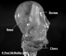
- Oedematous bursa (may be slightly enlarged, normal size or reduced in size depending on the stage), may have haemorrhages, rapidly proceeds to atrophy.
- Haemorrhages in skeletal muscle (especially on thighs).
- Dehydration.
- Swollen kidneys with urates.
Diagnosis
Clinical disease - History, lesions, histopathology.
Subclinical disease - A history of chicks with very low levels of maternal antibody (Fewer than 80% positive in the immunodifusion test at day old, Elisa vaccination date prediction < 7 days), subsequent diagnosis of 'immunosuppression diseases' (especially inclusion body hepatitis and gangrenous dermatitis) is highly suggestive. This may be confirmed by demonstrating severe atrophy of the bursa, especially if present prior to 20 days of age.
The normal weight of the bursa in broilers is about 0.3% of bodyweight, weights below 0.1% are highly suggestive. Other possible causes of early immunosuppression are severe mycotoxicosis and managment problems leading to severe stress.
Variants: There have been serious problems with early Gumboro disease in chicks with maternal immunity, especially in the Delmarva Peninsula in the USA. IBD viruses have been isolated and shown to have significant but not complete cross-protection. They are all sero-type 1. Serology: antibodies can be detected as early as 4-7 days after infection and these last for life. Tests used are mainly Elisa, (previously SN and DID). Half-life of maternally derived antibodies is 3.5- 4 days. Vaccination date prediction uses sera taken at day old and a mathematical formula to estimate the age when a target titre appropriate to vaccination will occur.
Differentiate clinical disease from: Infectious bronchitis (renal); Cryptosporidiosis of the bursa (rare); Coccidiosis; Haemorrhagic syndrome.
Treatment
No specific treatment is available. Use of a multivitamin supplement and facilitating access to water may help. Antibiotic medication may be indicated if secondary bacterial infection occurs.
Prevention
Vaccination, including passive protection via breeders, vaccination of progeny depending on virulence and age of challenge. In most countries breeders are immunised with a live vaccine at 6-8 weeks of age and then re-vaccinated with an oil-based inactivated vaccine at 18 weeks. A strong immunity follows field challenge. Immunity after a live vaccine can be poor if maternal antibody was still high at the time of vaccination.
When outbreaks do occur, biosecurity measures may be helpful in limiting the spread between sites, and tracing of contacts may indicate sites on which a more rebust vaccination programme is indicated.
 |
| Figure 23. This shows the anatomical relationship between the bursa, the rectum and the vent. This bursa is from an acutely affected broiler. It is enlarged, turgid and oedematous. |


