


|
Histopathology and Cytology of
Poultry Diseases By Ivan Dinev, DVM, PhD
|
ESCHERICHIA COLI INFECTIONS
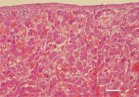
Fig. 1. Congestion (overfilling of blood vessels with red blood cells) of the liver as an initial manifestation of E. coli septicaemia in a broiler chicken. H/E, Bar = 40 µm.
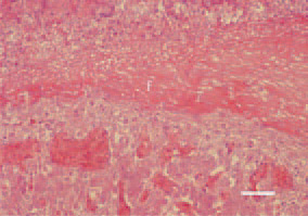
Fig. 2. Serofibrinous perihepatitis consequent to E. coli septicaemia in a broiler chicken. Huge fibrinous pseudomembrane (f), coating the liver surface. H/E, Bar = 50 µm
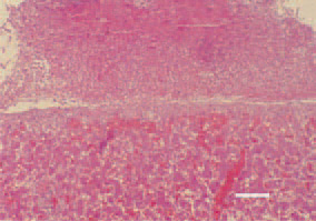
Fig. 3. Serofibrinous perihepatitis. Organization of pseudomembranous deposits. H/E, Bar = 50 µm.
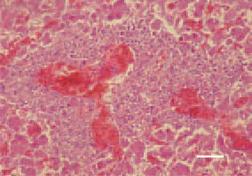
Fig. 4. Е. coli septicaemia. Massive perivascular liver necrosis. H/E, Bar = 30 µm.
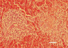
Fig. 5. Periarteriolar reaction, fibrinoid necrosis and congestion of the spleen in Е. coli septicaemia. H/E, Bar = 30 µm.
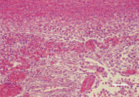
Fig. 6. Croupous pleuropneumonia - one of the commonest findings in Е. coli septicaemia of respiratory origin. H/E, Bar = 25 µm.
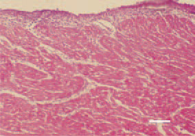
Fig. 7. Serofibrinous pericarditis and inflammatory oedema of the myocardium in Е. coli septicaemia. H/E, Bar = 30 µm.
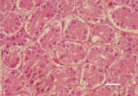
Fig. 8. Degenerative necrobiotic lesions in the epithelium of renal tubules. H/E, Bar = 25 µm.
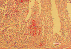
Fig. 9. Catarrhal haemorrhagic enteritis caused by an enterotoxigenic Е. coli strain. H/E, Bar = 100 µm.
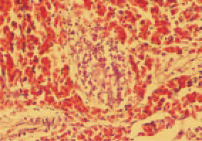
Fig. 10. Element of Fig. 9. Massive haemorrhages surrounding lymphatic clusters in the intestinal wall. H/E, Bar = 25 µm.
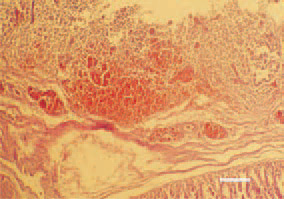
Fig. 11. Haemorrhages in the proventriculus mucous coat in colisepticaemia of enteric origin, secondary to necrotic enteritis. H/E, Bar = 100 µm.
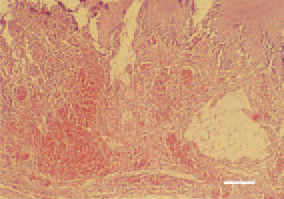
Fig. 12. Haemorrhages and cystic formations in the gizzard in colisepticaemia of enteric origin, secondary to necrotic enteritis. H/E, Bar = 40 µm.
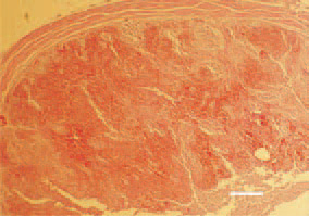
Fig. 13. Massive haemorrhages in the parenchyma of tonsila caecalis in colisepticaemia of enteric origin, secondary to necrotic enteritis. H/E, Bar = 50 µm.
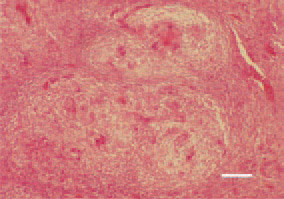
Fig 14. Coligranuloma (Hjarre’s disease). Conglomerates of granulomatous nodes against the intestinal wall. Central necrotic detritus and marked acidophilia. Single foreign body-type giant cells. H/E, Bar = 30 µm.
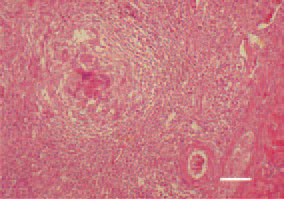
Fig 15. Coligranuloma (Hjarre’s disease). Coligranuloma, liver. H/E, Bar = 35 µm.
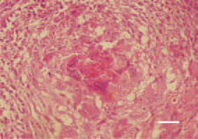
Fig. 16. Coligranuloma (Hjarre’s disease). Coligranuloma, liver. The typical heterophilic debris among the central necrotic detritus. H/E, Bar = 25 µm.
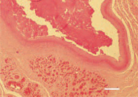
Fig. 17. Sternal bursitis. Filling of the sternal bursa with fibrinous caseous exudate. Peripheral outgrowth of fibrous tissue and inflammatory hyperaemia. H/E, Bar = 100 µm.