


|
Histopathology and Cytology of
Poultry Diseases By Ivan Dinev, DVM, PhD
|
RUPTURE OF THE GASTROCNEMIUS TENDON IN BROILER PARENTS
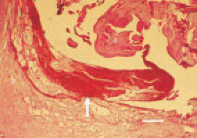
Fig. 1. Haemorrhagies (arrow) between the tendon (a) and the tendon sheath (b), H/E, Bar = 50 µm.
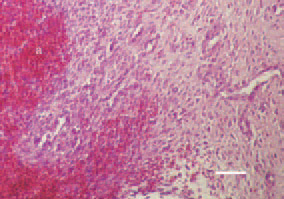
Fig. 2. Resolving haematoma (a) and fibrous tissue growth (b), H/E, Bar = 35 µm.
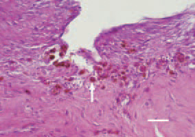
Fig. 3. Macrophages (siderocytes) (arrow) among the neocollagenous connective tissue, H/E, Bar = 30 µm.
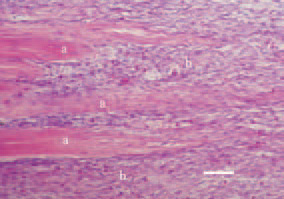
Fig. 4. The ends (a) of ruptured tendon, embraced by the newly grown connective tissue (b), H/E, Bar = 50 µm.
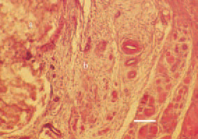
Fig. 5. Axial necrosis (а) of the gastrocnemius tendon; (b – intact tendinous tissue), H/E, Bar = 30 µm.
This book is protected by the copyright law.
The reproduction, imitation or distribution of the book in whole or in part, in any format (electronic, photocopies etc.) without the prior consent, in writing, of copyright holders is strictly prohibited.