


|
Histopathology and Cytology of
Poultry Diseases By Ivan Dinev, DVM, PhD
|
CHLAMYDIOSIS
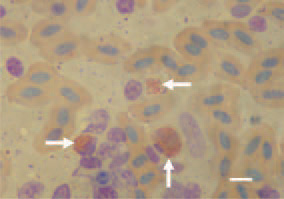
Fig. 1. Touch imprint preparation from the air sac of a duck. Intracytoplasmic red chlamydial elemental bodies. Giemsa stain, Bar = 10 µm.
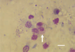
Fig. 2. Touch imprint preparation from the air sac of a duck. Macrophageal inflammatory cell response with intracellular inclusions. Diff Quik, Bar = 10 µm.
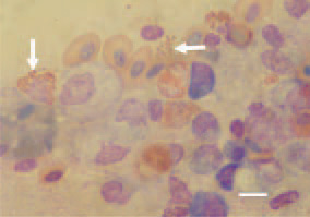
Fig. 3. Touch imprint preparation from the air sac of a duck. Multiple intracytoplasmic red chlamydial elemental bodies. Giemsa stain, Bar = 10 µm.
This book is protected by the copyright law.
The reproduction, imitation or distribution of the book in whole or in part, in any format (electronic, photocopies etc.) without the prior consent, in writing, of copyright holders is strictly prohibited.