


|
Histopathology and Cytology of
Poultry Diseases By Ivan Dinev, DVM, PhD
|
FATTY LIVER HAEMORRHAGIC SYNDROME
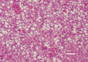
Fig. 1. Marked fatty dystrophy, vacuoles of a various size in hepatocytes and haemorrhages in the liver of a laying hen. H/E, Bar = 30 µm.
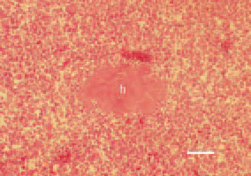
Fig. 2. Fatty liver haemorrhagic syndrome, laying hen. Organization of a haematoma (h) among the liver parenchyma. H/E, Bar = 40 µm.
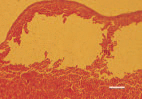
Fig. 3. Fatty liver haemorrhagic syndrome, laying hen. Resorbed subcapsular haematoma. H/E, Bar = 50 µm.
This book is protected by the copyright law.
The reproduction, imitation or distribution of the book in whole or in part, in any format (electronic, photocopies etc.) without the prior consent, in writing, of copyright holders is strictly prohibited.