


|
Histopathology and Cytology of
Poultry Diseases By Ivan Dinev, DVM, PhD
|
HAEMORRHAGIC ENTERITIS OF TURKEYS
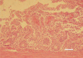
Fig. 1. Haemorrhagic desquamative catarrh of the mucosa in a 10-weekold turkey. H/E, Bar = 50 µm.
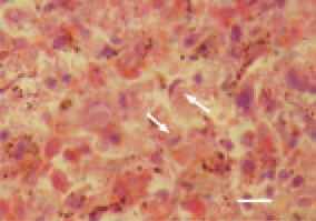
Fig. 2. A characteristic diagnostic finding is the detection of large, acidophilic, rarely basophilic intranuclear inclusion bodies in the reticuloendothelial cells of the spleen (arrows). The displaced condensed nuclear chromatin around the inclusion bodies often resembles a crescent. H/E, Bar = 10 µm.
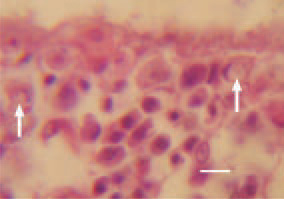
Fig. 3. Sometimes, similar inclusion bodies could be detected in the lamina propria of the intestinal mucosa (arrows). H/E, Bar = 10 µm.
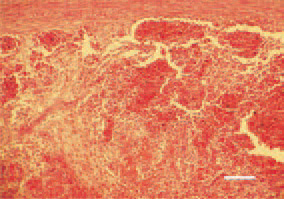
Fig. 4. In the spleen, hyperplasia of the white pulp, necroses, subcapsular and periarteriolar haemorrhages could be generally observed, consequently to a secondary E. coli septicaemia. H/E, Bar = 100 µm.
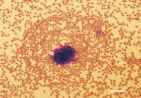
Fig. 5. The stained peripheral blood smears exhibit the typical picture of a septicaemia. H/E, Bar = 10 µm.