


|
Histopathology and Cytology of
Poultry Diseases By Ivan Dinev, DVM, PhD
|
AMYLOIDOSIS
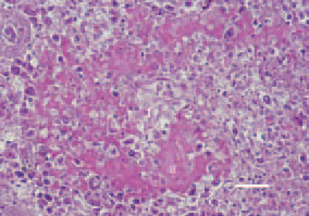
Fig. 1. Nodular form of amyloidosis, marble spleen disease, pheasant. Deposits of homogeneous amyloid masses (a) from the periphery toward the centre of white pulp follicles (f). Congo red, Bar = 25 µm.
This book is protected by the copyright law.
The reproduction, imitation or distribution of the book in whole or in part, in any format (electronic, photocopies etc.) without the prior consent, in writing, of copyright holders is strictly prohibited.