


|
Histopathology and Cytology of
Poultry Diseases By Ivan Dinev, DVM, PhD
|
ULCERATIVE ENTERITIS
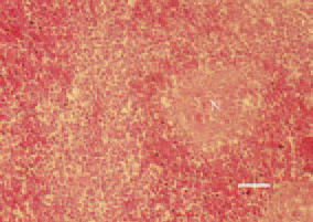
Fig. 1. Liver. Well- or poorly demarcated coagulative necroses (N) with minor inflammatory reaction and peripheral haemorrhagic zone. H/E, Bar = 40 µm.
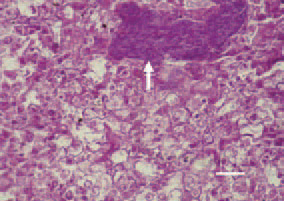
Fig. 2. Liver. The histological section reveals groups as well as intralesional UE organisms, spread among the parenchyma (arrow). H/E, Bar = 25 µm.
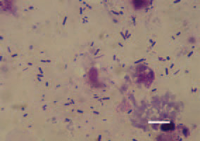
Fig. 3. Touch imprint preparation of intestinal content from a broiler chicken with UE. Multiple UE agents and desquamated epithelial cells from the necrotic mucous coat. Diff Quik, Bar = 10 µm.
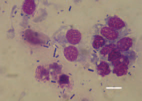
Fig. 4. Touch imprint preparation of a liver cross section. Typical UE microorganisms, straight or slightly curved rods with rounded tips. Diff Quik, Bar = 10 µm.
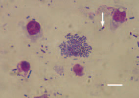
Fig. 5. Element of Fig. 4. A bacterium with characteristic subterminal spore (arrow). Diff Quik, Bar = 10 µm.