


|
Histopathology and Cytology of
Poultry Diseases By Ivan Dinev, DVM, PhD
|
MAREK’S DISEASE
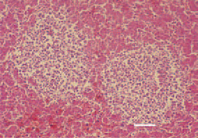
Fig. 1. Symmetrical neoplastic foci of pleomorphic cells, liver, hen. H/E, Bar = 50 µm.
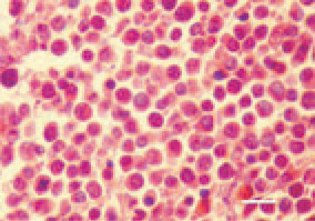
Fig. 2. Acute (visceral) form, liver, hen. Lymphomatous lesions, mainly consisting of lymphoblasts, and small to medium-sized lymphocytes. Among the proliferate, single plasmatic and reticular cells are outlined. H/E, Bar = 10 µm.
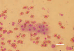
Fig. 3. Marek’s disease, pullet, touch imprint preparation of a liver crosssection. Lymphoid cells of a various size: lymphoblasts and lymphocytes. Diff Quik, Bar = 10 µm.
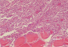
Fig. 4. Muscle tumour, Marek’s disease. Focal pleomorphic cell proliferation. H/E, Bar = 35 µm.
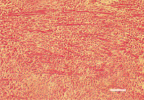
Fig. 5. Diffuse pleomorphic cell proliferation in the myocardium, resulting in atrophy of myofibrils. H/E, Bar = 35 µm.
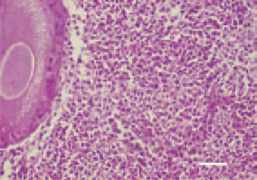
Fig. 6. Pleomorphic cell proliferation, ovary, hen. H/E, Bar = 25 µm.
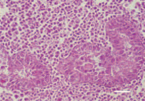
Fig. 7. Intertubular, pleomorphic cell proliferation, testis, cock. H/E, Bar = 30 µm.
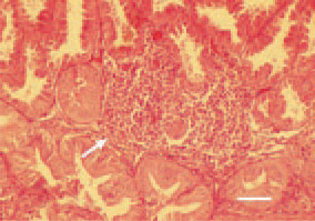
Fig. 8. Diffuse pleomorphic cell proliferation in the proventriculus mucous coat (arrow), hen. Compression and atrophy of glandular acini. H/E, Bar = 40 µm.
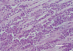
Fig. 9. Peripheral nerve. A-type lesion (neoplastic type), marked lymphoid cell proliferation, absence of oedema. H/E, Bar = 25 µm.
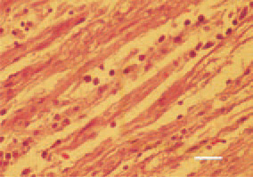
Fig. 10. Peripheral nerve, B-type lesion (mostly inflammatory type). Interneuritic inflammatory oedema and slight to moderate proliferation of lymphocytes and plasmatic cells, rarely lymphoblasts. The mild version of B-type lesions is called C-type. H/E, Bar = 25 µm.
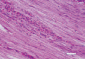
Fig. 11. Case of focal myelocytic proliferation of the sciatic nerve, hen. Bar = 25 µm.
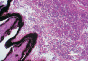
Fig. 12. Lymphoid cell proliferations in the iris and ciliary muscles in the ocular form of Marek’s disease. H/E, Bar = 50 µm.