


|
Histopathology and Cytology of
Poultry Diseases By Ivan Dinev, DVM, PhD
|
GOUT
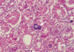
Fig. 1. Visceral gout. Transverse crosssection, kidney, chicken. Bluish-purple urate deposits in the lumen of renal tubules forming urate cylinders (arrows). H/E, Bar = 25 µm.
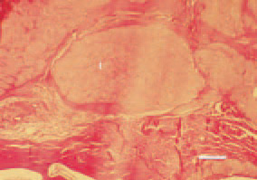
Fig. 2. Articular gout, transverse cross-section, phalange, cock. Amorphous masses (urates) in the periarticular tissue, forming tophi (t), surrounded by fibrous tissue. H/E, Bar = 50 µm.
This book is protected by the copyright law.
The reproduction, imitation or distribution of the book in whole or in part, in any format (electronic, photocopies etc.) without the prior consent, in writing, of copyright holders is strictly prohibited.