


|
Histopathology and Cytology of
Poultry Diseases By Ivan Dinev, DVM, PhD
|
STREPTOCOCCOSIS
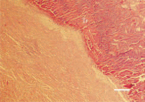
Fig. 1. Valve thromboendocarditis. Massive thrombotic masses (Tr) coating the mitral valve in the left heart side of a duck. It is generally associated with Streptococcus zooepidemicus. H/E, Bar = 40 µm.

Fig. 2. Streptococcosis, duck. A mixed wall thrombus (Tr) partially occluding the left atrioventricular opening. H/E, Bar = 50 µm.
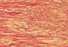
Fig. 3. Myocardial infarction (I) as a result of thromboemboly. Focal degenerative necrobiotic lesions and beginning of organization, appearance of macrophages. H/E, Bar = 25 µm.
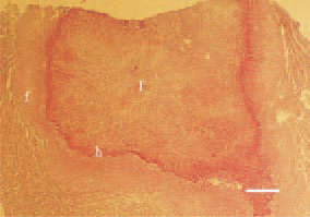
Fig. 4. Widespread anaemic infarct (I) in the spleen of a duck. A peripherally reactive marginal zone of hyperaemia (h), necrotic heterophils and early fibroblastosis (f). The lesion is clearly delineated by the adjacent healthy parenchyma and is generally resulting from a portal venous thrombosis. H/E, Bar = 100 µm.