|
Histopathology and Cytology of
Poultry Diseases By Ivan Dinev, DVM, PhD
|
DEEP PECTORAL MYOPATHY
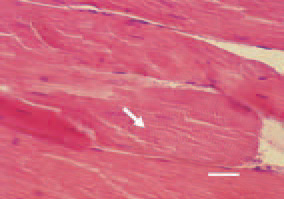
Fig. 1. The condition is resulting from ischaemic necrosis due to inadequate blood supply of deep pectoral muscle groups of a various size. Initial stage of DPM, characterized with discoid necrosis (arrow) of muscle fibres, manifested by separation of single sarcomeres. H/Е, bar = 25 µm.
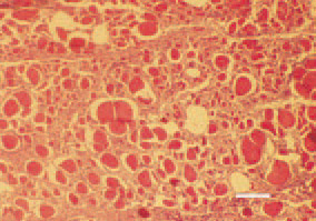
Fig. 2. Histologically, the altered muscle fibres appear enlarged at a various extent, distinctly eosinophilic with rhexic or absent nuclei. H/E, Bar = 40 µm.
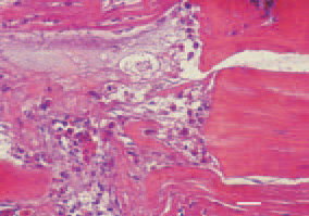
Fig. 3. Degenerative necrobiotic changes from the Zenker’s necrosis type, enhanced acidophilia, heterophil leukocytes and macrophages among the dead muscle tissue. H/Е, bar = 35 µm.
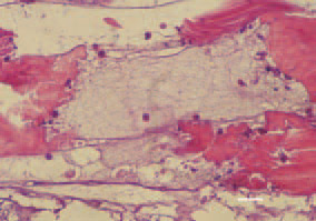
Fig. 4. Mucoid degeneration and a distinct border between the affected and intact muscle tissue areas. H/Е, bar = 35 µm.
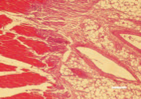
Fig. 5. At a later stage, a substitution of atrophied fibres by adipose tissue could be detected. H/E, Bar = 40 µm.
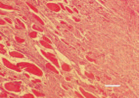
Fig. 6. A late (chronic) lesion. The place of necrotic musculature is occupied by fibrous tissue. H/Е, bar = 50 µm.


