


|
Histopathology and Cytology of
Poultry Diseases By Ivan Dinev, DVM, PhD
|
RICKETS
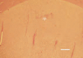
Fig. 1. Histopathological lesions in rickets due to calcium and vitamin D deficiency. The proliferative zone of the growth plate is widened, irregular and poorly vascularized. Only a small zone of hypertrophic cartilage and reduced calcification is present. H/E, Bar = 40 µm.
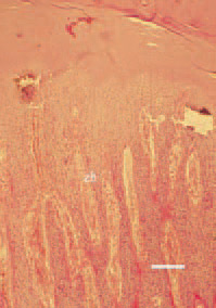
Fig. 2. Histopathological lesions in rickets due to phosphate deficiency or excessive calcium levels. The hypertrophic zone (zh) of the growth plate is increased, noncalcified, but normally vascularized by metaphyseal blood vessels. H/E, Bar = 40 µm.
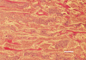
Fig. 3. A hypertrophic zone, consisting of multiple nonmineralized trabeculae of cartilage in a case of vitamin D deficiency. H/E, Bar = 35 µm.
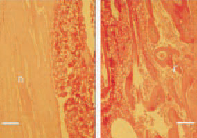
Fig. 4. In cases of prolonged calcium deficiency, a removal of calcium from the skeleton does occur. Consequently, thinning of cortices of long bones is resulting. Longitudinal cross-section, femurs, 40-day-old broiler chickens. Left: normal thickness of the bone wall (n). Right: extreme thinning of the cortex (r) due to demineralization in a broiler chicken. H/E, Bar = 50 µm.
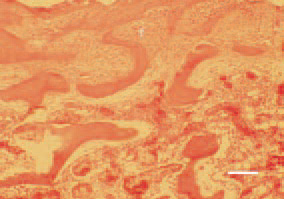
Fig. 5. Sagittal cross-section of the proximal femur in a broiler chicken, case of calcium deficiency. Replacement of bone substance by a newly grown fibrous (f) tissue. H/E, Bar = 30 µm.
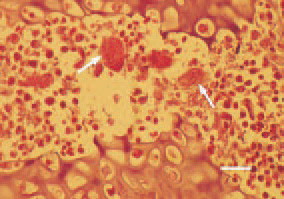
Fig. 6. The detection of osteoclasts (arrows) in some cases of calcium deficiency rickets resembles the findings in osteoporosis of laying hens. H/E, Bar = 25 µm.
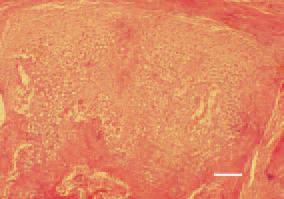
Fig. 7. Tarsometatarsal bone, 35-dayold broiler chicken, longitudinal crosssection. Growth of subperiosteal abnormal masses of hypertrophic cartilage, resulting in extensive bone thickening looking like osteopetrosis. H/E, Bar = 35 µm.