


|
Histopathology and Cytology of
Poultry Diseases By Ivan Dinev, DVM, PhD
|
SPIROCHAETOSIS
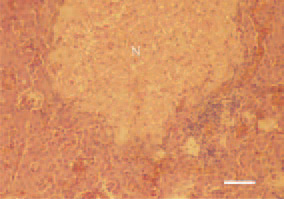
Fig. 1. Liver, hen. Well-demarcated coagulative necrosis (N) against the surrounding parenchyma. Marked peripheral reaction through inflammatory cell proliferate and hyperaemia. H/E, Bar = 50 µm.
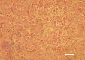
Fig. 2. Reactive necrosis (N), liver, hen. Multiple eosinophilic granulocytes among the necrotic focus and perivascular inflammatory cell clusters. H/E, Bar = 35 µm.
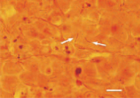
Fig. 3. B. anserina, of a filiform helical shape, mucronate at the apexes (arrows) among the hepatic cells. Liver cross-section, Levaditi staining, Bar = 25 µm.
This book is protected by the copyright law.
The reproduction, imitation or distribution of the book in whole or in part, in any format (electronic, photocopies etc.) without the prior consent, in writing, of copyright holders is strictly prohibited.