


|
Histopathology and Cytology of
Poultry Diseases By Ivan Dinev, DVM, PhD
|
PULMONARY HYPERTENSION (ASCITES) SYNDROME IN BROILER CHICKENS
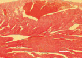
Fig. 1. Separation of intermyocardial fibres by a moderate to extensive oedema of loose connective tissue. Subepicardial and myocardial haemorrhages of a various size. H/E, Bar = 50 µm.
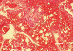
Fig. 2. Lung. Hyperaemia, haemorrhages and oedema. A possible finding is the appearance of of bone and cartilage foci (co) among the pulmonary parenchyma. H/E, Bar = 30 µm.
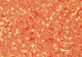
Fig. 3. Liver sinusoids are enlarged and overfilled with blood. Supplementary hepatic findings are the oedema and atrophic degenerative lesions. H/E, Bar = 50 µm.
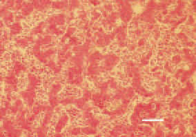
Fig. 4. Compression atrophy of liver parenchyma secondary to congested sinusoids resulting from the passive venous hyperaemia. H/E, Bar = 30 µm.
This book is protected by the copyright law.
The reproduction, imitation or distribution of the book in whole or in part, in any format (electronic, photocopies etc.) without the prior consent, in writing, of copyright holders is strictly prohibited.