


|
Histopathology and Cytology of
Poultry Diseases By Ivan Dinev, DVM, PhD
|
ASPERGILLOSIS
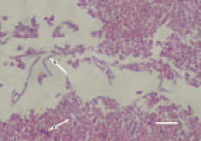
Fig. 1. In acute aspergillosis, fungal spores (arrow – a) and grown hyphae (arrow – b) could be observed among the inflammatory necrotic masses. H/E, Bar = 30 µm.
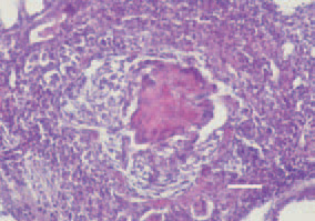
Fig. 2. In the nodular form, the characterisitic Aspergillus granuloma structure is seen. A central necrosis, surrounded by foreign-body giant cells arranged in a wreath. H/E, Bar = 30 µm.
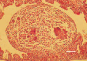
Fig. 3. Aspergillus granuloma to the highly corrugated mucosa of a parabronchus, lung, chicken. H/E, Bar = 25 µm.
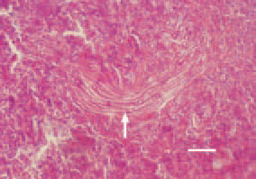
Fig. 4. Aspergillosis, liver, chicken. Aspergillus mycelium outgrowth (arrow). H/E, Bar = 50 µm.
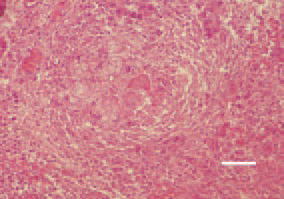
Fig. 5. A shaped Aspergillus granuloma in the liver of a chicken. H/E, Bar = 35 µm.
This book is protected by the copyright law.
The reproduction, imitation or distribution of the book in whole or in part, in any format (electronic, photocopies etc.) without the prior consent, in writing, of copyright holders is strictly prohibited.