


|
Histopathology and Cytology of
Poultry Diseases By Ivan Dinev, DVM, PhD
|
HISTOMONIASIS
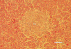
Fig. 1. Histomoniasis, liver, turkey poult. Coagulative necrotic foci (N), haemorrhagically demarcated from the adjacent liver parenchyma. H/E, Bar = 40 µm.
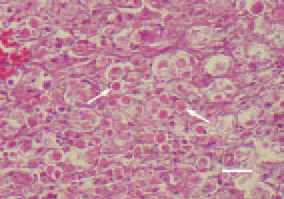
Fig. 2. Multiple oval-shaped histomonads (arrows) in a liver tissue cross-section, pheasant. H/E, Bar = 25 µm.
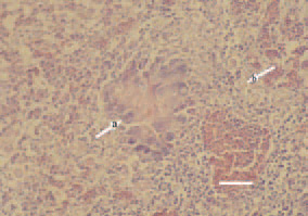
Fig. 3. Histomoniasis, liver, turkey poult. Initial organization of a necrotic focus in the liver. A wreath of foreign-body giant cells (arrow – a) in the periphery of necrotic area. Parasites (arrow – b) are also seen on the tissue cross-section. H/E, Bar = 35 µm.
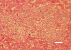
Fig. 4. Regenerative reparative processes at a later stage of the organization of a hepatic necrotic focus in a turkey poult. Histomonas forms (arrows) are still visible. H/E, Bar = 40 µm.
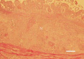
Fig. 5. Histomoniasis, caecum – transverse cross-section, turkey poult. Coagulative necrosis (N) affecting the mucosa and submucosa of the intestinal wall. H/E, Bar = 100 µm.