


|
Histopathology and Cytology of
Poultry Diseases By Ivan Dinev, DVM, PhD
|
SALMONELLOSES
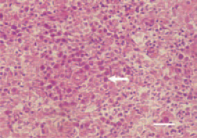
Fig. 1. Pullorum disease. Perivascular proliferative inflammatory focus in the liver, consisting mainly of histiocytes and single epitheloid cells (arrow). H/E, Bar = 25 µm.
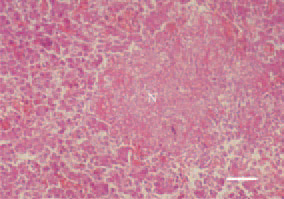
Fig. 2. Fowl typhoid. Areactive fibrinoid necrosis (N) in the liver. H/E, Bar = 40 µm.
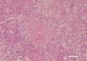
Fig. 3. Fowl typhoid. Periarteriolar fibrinoid necrosis (N) in the spleen. Marked cell reaction in the periphery of the necrotic foci involving lymphocytes, histiocytes and single granulocytes. H/E, Bar = 50 µm.
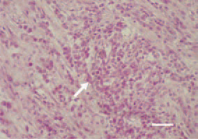
Fig. 4. Fowl typhoid, hen. Mononuclear inflammatory cell proliferate in the myocardium (arrow). H/E, Bar = 30 µm.
This book is protected by the copyright law.
The reproduction, imitation or distribution of the book in whole or in part, in any format (electronic, photocopies etc.) without the prior consent, in writing, of copyright holders is strictly prohibited.