


|
Histopathology and Cytology of
Poultry Diseases By Ivan Dinev, DVM, PhD
|
VITAMIN E DEFICIENCY
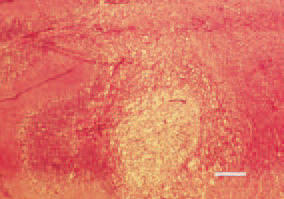
Fig. 1. Histologically, colliquation necroses are observed, mainly in the white brain substance of corpus medularis, appearing as brighter foci, extensively vacuolized and with a reticular structure. H/E, Bar = 30 µm.
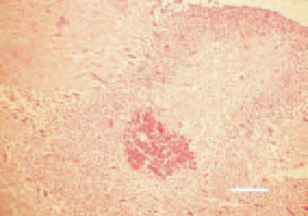
Fig. 2. Haemorrhages of a various size are observed among the necrotic tissue. H/E, Bar = 35 µm.
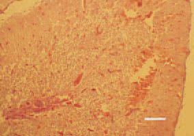
Fig. 3. Haemorrhages and multiple thrombosed blood vessels in the cerebelum. H/E, Bar = 100 µm.
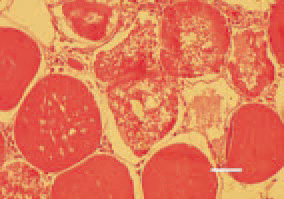
Fig. 4. Muscular dystrophy, pectoral musculature, hen. Hyaline degeneration of muscle fibres (waxy degeneration). Swol len homogeneous muscle fibres, having lost their striated pattern, with increased eosinophilia. Centrally, necrobiotic lesions resulting in muscle cell degradation (Zenker’s necrosis). H/E, Bar = 25 µm.
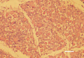
Fig. 5. Late stage of muscular dystrophy, thigh muscle, chicken. Marked acidophilia of muscle cells and beginning of regenerative reparative processes, organization. H/E, Bar = 35 µm.