|
Histopathology and Cytology of
Poultry Diseases By Ivan Dinev, DVM, PhD
|
IONOPHORE TOXICITY CHICKENS
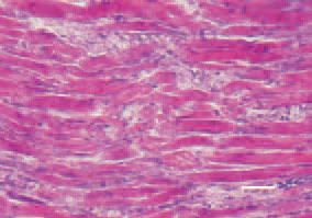
Fig. 1. & Fig. 2. Transverse/londitudinal cross-section, thigh muscle, chicken, after intoxication with high doses maduramycin. Enhanced eosinophilia and denenerative necrobiotic lesions of muscle fibres. H/E, Bar = 40 µm.
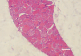
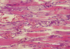
Fig. 3. Longitudinal cross-section, thigh muscle, chicken, after intoxication with high doses maduramycin. Appearance of macrophages and initial organization of necrotic detritus occuring after muscle fibre breakdown. H/E, Bar = 50 µm.
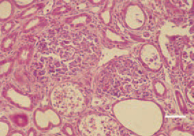
Fig. 4. Inflammatory, degenerative necrobiotic lesions and urate deposits in renal tubules. H/E, Bar = 25 µm.
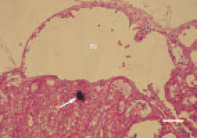
Fig. 5. Urate cylinders (arrow) and retention cysts (rc) in kidneys. H/E, Bar = 50 µm.
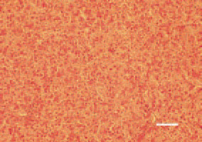
Fig. 6. Degenerative necrobiotic lesions of the liver. H/E, Bar = 50 µm.
This book is protected by the copyright law.
The reproduction, imitation or distribution of the book in whole or in part, in any format (electronic, photocopies etc.) without the prior consent, in writing, of copyright holders is strictly prohibited.


