


|
Histopathology and Cytology of
Poultry Diseases By Ivan Dinev, DVM, PhD
|
AVIAN TUBERCULOSIS
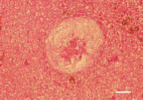
Fig. 1. Tubercle, liver, ostrich. Centrally, a cluster of epitheloid cells (that are fusiform in birds), radially directed to the centre of the granuloma, is visible. Appearance of foreign bodytype giant cells and initial capsulation. H/E, Bar = 50 µm.
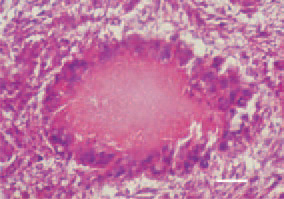
Fig. 2. Tubercle, liver, hen. Centrally, a caseous necrosis of gathered epitheloid cells, forming the basic mass of the granuloma. Peripherally, foreign body-type giant cells, arranged one by the other in a wreath-like fashion. H/E, Bar = 35 µm.
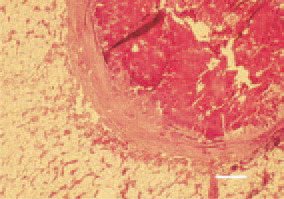
Fig. 3. Tubercle, bone marrow, ostrich. Caseous necrosis and capsulation of the granuloma. H/E, Bar = 25 µm.
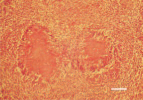
Fig. 4. Symmetrical, caseous necrotic foci in the intestinal wall in a hen. Peripherally, foreign body-type giant cells. H/E, Bar = 40 µm.
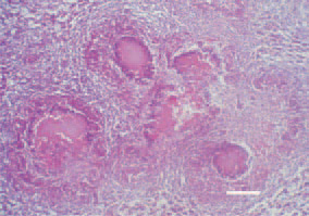
Fig. 5. Tubercle, liver, hen. A characteristic finding in avian tuberculosis are conglomerate tubercles, formed by several nodules having merged into a common mass. H/E, Bar = 50 µm.