


|
Histopathology and Cytology of
Poultry Diseases By Ivan Dinev, DVM, PhD
|
NECROTIC ENTERITIS
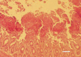
Fig. 1. Necrosis of small intestinal mucous coat. The affected mucosal layer is altered into necrotic detritus, part of which protrudes into the intestinal lumen. H/E, Bar = 40 µm.
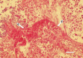
Fig. 2. Simultaneous presence of Clostridium organisms (arrow - с) and Eimeria developmental forms (arrow - е) among the necrotic small intestinal mucosa in a broiler chicken. H/E, Bar = 30 µm.
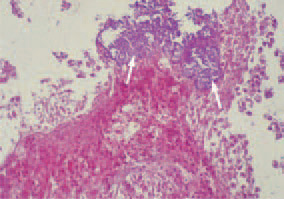
Fig. 3. Element of Fig. 2. Massive clostridial colonization to the surface of small intestinal mucosa (arrows). H/E, Bar = 35 µm.
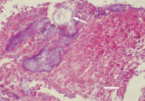
Fig. 4. Multiple clostridiae among the necrotic intestinal mucosa and formation of a cavity (arrow) as a result of gas production. H/E, Bar = 25 µm.
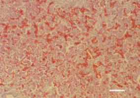
Fig. 5. Necrotic enteritis in a broiler chicken. Severe liver congestion. H/E, Bar = 100 µm.
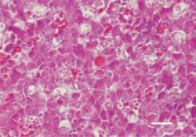
Fig. 6. Necrotic enteritis in a broiler chicken. Liver, activated macrophages and enhanced phagocytosis of erythrocytes. H/E, Bar = 35 µm.