


|
Histopathology and Cytology of
Poultry Diseases By Ivan Dinev, DVM, PhD
|
VITAMIN A DEFICIENCY
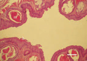
Fig. 1. Transverse cross-section of the oesophagus, hen. In some instances, miliary grey-whitish thick nodules, prominating over the surface of the buccal mucosa, the oesophagus and the crop, could be observed. They are result of the occurring hyperkeratinization (h) and metaplasia (m) of the glandular epithelium. H/E, Bar = 35 µm.
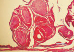
Fig. 2. Hypovitaminosis А, pigeon. Hyperkeratinization of follicles of B. Fabricii mucous coat. H/E, Bar = 35 µm.
This book is protected by the copyright law.
The reproduction, imitation or distribution of the book in whole or in part, in any format (electronic, photocopies etc.) without the prior consent, in writing, of copyright holders is strictly prohibited.